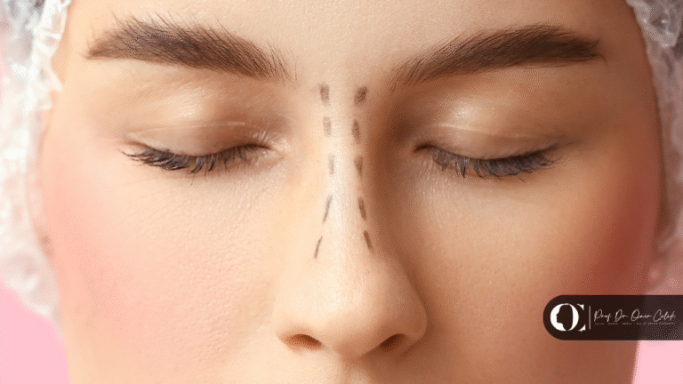


Rib cartilage is a support material used when the cartilage inside the nose has run out or is insufficient. It is most often chosen in revision operations, severe deformities and cases where the nasal bridge needs to be raised significantly. Warping, the most feared problem, was reported at a rate of 3% for autologous rib in a review of 2,322 patients. All the grafts the operation needs can be obtained from a single site.
For many patients, the sentence “cartilage will be taken from the rib” causes more anxiety than the operation itself.
The anxiety has two sources: the fact that an incision will be made on the chest, and the fear that the cartilage will warp over time.
This article addresses both with figures — and the picture that emerges is quite different from the common perception.
When is rib cartilage needed?
Rib is not the first choice. It comes last in the order of preference.
The cartilage hierarchy
| Source | Advantage | Limitation |
|---|---|---|
| Septum (inside the nose) | Straight, strong, no additional incision | Limited amount; may have been used in a previous operation |
| Ear (concha) | Easy to access | Naturally curved; not suitable for dorsal support |
| Rib | Plentiful, strong structural support | Requires a separate incision site |
If the septum is sufficient, rib is not needed. The problem is that the septum is not always sufficient.
Situations where rib comes to the fore
- Revision rhinoplasty — if the septum was used or weakened in a previous operation
- Reconstruction after severe deformity or trauma
- Cases where the nasal bridge needs to be raised significantly
- Nasal collapse — conditions in which structural support has been largely lost
What these situations have in common: the aim here is not to shape but to build a load-bearing framework. And this requires robust, plentiful and straight material.
This is also where rib’s most practical advantage lies: all the grafts the operation needs can be obtained from a single site.
Nasal collapse itself and its causes are covered in a separate article; this article focuses on the material used in the repair.
The most feared issue: warping
This is the known weak point of rib cartilage. But the measured rate is well below what is commonly perceived.
What is warping?
Cartilage tissue contains natural internal tension forces. When cartilage is cut, the balance of these forces is disturbed and the piece tends to curl over time.
In the nose, this can appear as an unwanted curve on the bridge or at the tip.
What do the figures say?
A systematic review and meta-analysis covering 25 studies and 2,322 patients reported the following pooled rates for three different types of rib cartilage:
| Graft type | Warping | Infection | Revision |
|---|---|---|---|
| Autologous rib (patient’s own) | 3% | 2% | 7% |
| Irradiated homologous (donor, sterilised) | 1% | 1% | 7% |
| Fresh frozen (donor) | 1% | 1% | 1% |
An instructive asymmetry in the table
What deserves attention is this:
The most feared problem is 3%. But the reoperation rate is 7%.
In other words, warping is not the main reason these operations are redone. The need for revision mostly arises from other causes — such as a mismatch with aesthetic expectations, asymmetry or breathing problems.
This matters for directing the patient’s concern to the right place: rather than focusing on a single risk, it is more meaningful to assess the operation as a whole.
The review’s honest gap: resorption
One of the questions patients ask most often is “will the cartilage dissolve over time?”
The review could not give a pooled rate for this. The reason is stated clearly: resorption was reported inconsistently and has no standard definition.
This does not mean the answer is “no, it does not dissolve”. It means the literature has not yet produced a clean answer to this question.
What reduces warping?
The low rate is no accident; it is the result of known technical principles.
The balanced cross-section principle
The principle described by Gibson and Davis in 1958 is this: if the distorting forces are balanced along the cartilage, the graft does not warp. The researchers followed 46 grafts prepared with this method for three years.
In practice, this means carving the cartilage symmetrically.
The central portion is used
Grafts are typically prepared from the central part of the rib. Peripheral portions and shavings are mostly not used, because of their tendency to warp.
This also limits the answer to “how much graft can one rib provide”: not all of the harvested piece is usable.
The oblique split technique
In this method, described by Taştan and colleagues in 2013, the cartilage is cut at a specific angle to its long axis to obtain the longest possible grafts. Preserving the outer cortex contributes to stability.
The team that described the method reported no warping during follow-up.
An honest note: the data on this technique are largely based on clinical observation; large studies reporting comparative rates are limited.
A fact to keep in mind
The sources also state clearly that warping can still occur despite careful symmetrical carving, especially in structural grafts.
In other words, technique reduces the risk — it does not eliminate it.
Calcification: a problem thought to be age-related, but not only
This section challenges a common assumption.
What is calcification, and why does it matter?
It is the hardening of cartilage tissue through calcium deposition. Calcified cartilage is unsuitable for carving; it is brittle and does not take the desired shape.
For this reason, cartilage quality needs to be assessed in advance in every patient for whom rib is planned.
The common assumption and the reality
The common expectation is that calcification is related to older age.
However, according to the data reported, calcification was found in 22.5% of adolescent female patients, and it can begin as early as age 14.
Age and sex are reported as the factors most closely linked to cartilage quality — but being young does not mean the cartilage will be free of calcification.
How is it assessed?
- Rib X-ray — practical, low cost, low radiation
- Computed tomography — shows the calcification pattern in most detail, but at higher cost and radiation dose
- Probing the cartilage with a fine needle — during surgery
The source states that a simple rib X-ray is usually sufficient in practice.
The threshold
Calcification above 25% is considered significant. Mild calcification confined to the edges usually causes no problems, whereas central or granular moderate calcification is generally considered unsuitable.
Which rib is used?
It is not a random choice; shape and safety are decisive.
| Rib | Characteristics |
|---|---|
| 7th rib (right) | Generally preferred: anatomically the safest position, suitable length and width |
| 6th rib | Good depth and width, but a shorter straight section with a slight curve; care is needed in patients with breast implants |
| 8th rib | Narrow; not suitable for dorsal grafts |
| 11th and 12th ribs | Floating ribs; because they are naturally straight, they need less carving and warp less |
The last row is notable: the reason some surgeons prefer the floating ribs is that they reduce the warping problem from the outset.
The chest side: scar, pain, recovery
This is the part patients are told least about, yet are most curious about.
Incision and scar
- The incision is about 2 cm, horizontal and placed in a skin crease
- Closure is done in four layers: muscle, fascia, subcutaneous tissue and skin
- Multilayer closure reduces the tension caused by upper-body movement
- The chest wound is compressed with a light dressing for 3 days; a drain is usually not needed
- Stitches are removed after one week
- Silicone scar tape can be used for about two months
In patients with a history of keloid or hypertrophic scarring, triamcinolone may be applied to the area as a preventive measure.
Pain
Pain in the chest area is the most frequently reported complaint.
| Period | What to expect |
|---|---|
| Days 3–4 | Pain peaks |
| First week | Gradual decrease |
| ~3 months | Time to full resolution reported in the literature |
With a muscle-sparing approach, pain is reported to peak on days 3–4 and to decrease within about a week. The literature reports a peak in the first week followed by gradual easing over about three months.
Pain persisting for months is rare but possible — especially in patients who have had cartilage harvested more than once.
Return to activity
- The next day: light activities such as walking and sitting are encouraged
- About 3 weeks: strenuous activities such as running, weight training or playing an instrument
The timetable for the nose works differently; it is covered in a separate article.
Risk of pneumothorax
Injury to the lining of the lung is a known but rare complication of rib harvest.
The donor-site complications reported in the systematic review include pain, hypertrophic scar or keloid, infection and, rarely, pneumothorax. However, because these complications were not consistently defined and collected, no pooled rate could be calculated.
Practical protection is described as follows: staying in the midline during needle probing and not approaching the pleura. A routine post-operative chest X-ray is recommended until the surgeon has gained experience with this procedure.
Alternatives
Rib is not the only option.
Donor cartilage
The two options in the table:
- Irradiated homologous rib (IHCC): warping 1%, infection 1%, revision 7%
- Fresh frozen rib (FFCC): warping 1%, infection 1%, revision 1%
The advantage is that no incision is made on the chest and operating time is shorter. The review states that these options showed a safety profile comparable to autologous rib in terms of warping and infection.
An important limitation: the authors stress that the lower revision rate of FFCC should be interpreted with caution because of heterogeneity and design differences between studies. In addition, the supply of donor tissue and the related regulations vary from country to country.
Ear cartilage
Because of its natural curvature it is not suitable for dorsal support, but it can be used at the tip and for small grafts.
How is the choice made?
The review’s concluding sentence is clear on this: graft selection should be made individually, according to the patient’s anatomy, available resources and the surgeon’s experience — not according to long-held assumptions about safety.
Points to discuss at the assessment
- Is there usable cartilage left in the septum? Is rib really necessary, or can it be solved with less intervention?
- Has cartilage quality been assessed? Has imaging been done for calcification?
- Which rib is planned?
- Is donor cartilage an option? If so, what are its advantages and limitations?
- Where will the chest scar be, and how large?
- Is there a history of keloid or hypertrophic scarring?
- What is the recovery timetable? For both the nose and the chest.
Summary
Rib is the last step, not the first choice. It is not used if the septum is sufficient. It comes to the fore in revision operations, severe deformities, post-traumatic reconstruction and cases where the nasal bridge needs to be raised significantly. Its most practical advantage is that all grafts can be obtained from a single site.
The fear of warping is greater than the measured rate. In the review of 2,322 patients, warping with autologous rib was 3% and infection 2%. The revision rate, by contrast, was 7% — so warping is not the main reason for reoperation. The resorption rate could not be pooled because of inconsistent reporting; the literature does not yet have a clean answer to this question.
Calcification is not only a problem of older age. It was found in 22.5% of adolescent female patients and can begin as early as age 14. Cartilage quality therefore cannot be assumed from age; it needs to be assessed in advance.
The chest side is a recovery process too. An incision of about 2 cm, pain peaking on days 3–4, light activity the next day and strenuous activity after about 3 weeks.
Which material is suitable for your situation can be determined when your previous surgical history, the current state of your cartilage and the scope of the planned repair are assessed together. This assessment requires a medical examination.
Frequently Asked Questions
When is rib cartilage needed?
It is most often used in revision rhinoplasty when the septal cartilage has run out or been weakened, in reconstruction after severe deformity or trauma, in nasal collapse and in cases where the nasal bridge needs to be raised significantly. If the septum is sufficient, rib is not needed.
Does rib cartilage warp?
It can, but the measured rate is below the common perception. In a systematic review covering 25 studies and 2,322 patients, the pooled warping rate for autologous rib was reported as 3%. Symmetrical carving based on the balanced cross-section principle reduces the risk but does not eliminate it completely.
Will the cartilage dissolve over time?
The literature does not have a clear answer to this question. The same systematic review states openly that it could not calculate a pooled rate because resorption was reported inconsistently and has no standard definition.
Will there be a scar on my chest?
The incision is about 2 cm long, horizontal and placed in a skin crease. A four-layer closure is used to reduce tension. Stitches are removed after one week, and silicone scar tape can be applied for about two months. In people with a history of keloids, preventive treatments may be considered.
How long does the chest pain last?
Pain is the most frequently reported complaint. With a muscle-sparing approach, it is reported to peak on days 3–4 and decrease within about a week; the literature describes a peak in the first week followed by gradual easing over about three months. Pain lasting for months is rare but possible.
When can I return to sport?
For the chest: light activities such as walking and sitting are encouraged from the next day; strenuous activities such as running and weight training after about three weeks. The nose has its own timetable, which is assessed separately.
What is the risk of pneumothorax?
Injury to the lining of the lung is a known but rare complication. In the systematic review, no pooled rate could be calculated because donor-site complications were not consistently defined. For protection, staying in the midline and not approaching the pleura is recommended, together with a routine post-operative chest X-ray until experience has been gained.
Which rib is used?
The choice is usually made among the 6th to 8th ribs; the right 7th rib is generally preferred because of its anatomical safety and suitable length and width. The 8th rib is narrow and therefore not suitable for dorsal grafts. Some surgeons prefer the floating 11th and 12th ribs because they are naturally straight.
I am young — will my cartilage be sound?
Age alone is not decisive. In one reported study, calcification was found in 22.5% of adolescent female patients, and it was reported that this can begin as early as age 14. For this reason, it is recommended to assess cartilage quality in advance with a rib X-ray or CT scan.
Can donor cartilage be used instead of my own rib?
Irradiated homologous and fresh frozen rib options are available; the review states that they show a profile comparable to autologous rib in terms of warping and infection. The authors stress that the difference in revision rates should be interpreted with caution because of heterogeneity between studies. Availability varies by country.
Prof. Dr. Öner Çelik — Specialist in Otorhinolaryngology and Head and Neck Surgery. About
This content is for information only, does not constitute medical advice and does not replace a medical examination. The rates given are pooled results of published studies; individual outcomes vary with anatomy, previous surgical history and clinical situation. For your own situation, please consult a doctor.
References
- Wee JH et al. Complications associated with autologous rib cartilage use in rhinoplasty: a meta-analysis. JAMA Facial Plast Surg, 2015. pubmed.ncbi.nlm.nih.gov
This content is for information only and does not replace medical advice. Every medical procedure requires an individual assessment and may carry risks. Results vary from person to person.