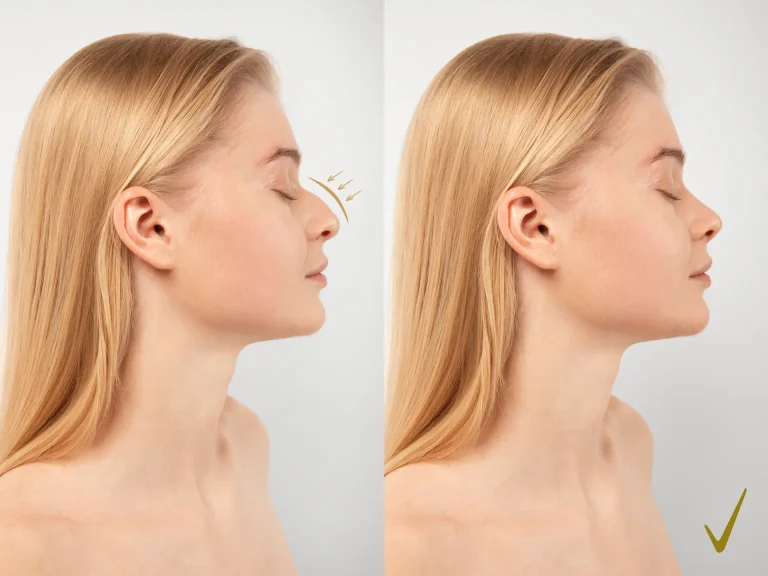


A saddle nose is a structural loss that appears as a collapse of the nasal bridge and is almost always accompanied by breathing problems. The underlying mechanism is the loss of the septal support that holds up the nose. Before any correction is planned, the cause must be investigated — because some causes require medical treatment before surgery. In mild and moderate cases, results are markedly better than in advanced cases.
A saddle nose is recognised by its appearance in the mirror: the bridge has collapsed, the profile is concave and the tip appears to turn upwards.
But this picture has two features, and both are often overlooked.
First: it is almost never only about appearance. The structure that collapses is also the structure that keeps the airway open.
Second: it is not a disease but a consequence. And correcting it without investigating the cause is not the right approach.
In this article I explain why it develops, which causes need a medical assessment first, and how reconstruction is planned.
The mechanism: loss of the supporting column
The height of the nasal bridge is carried by a structure beneath the visible part.
The front and upper portion of the septum forms a support known in the literature as the L-strut. This structure is the column that carries the bridge and the tip of the nose.
The definition in the literature is clear: a saddle nose is the loss of dorsal height resulting from insufficiency of the septal support column or the middle vault.
In other words, the problem is not in the skin, the fat or the muscle — it is in the framework.
That is why the solution is not a filler or a superficial procedure either, but rebuilding the structure.
Why is breathing affected too?
Because the same structure does two jobs at once.
The septum and the middle vault form the walls of the internal nasal valve, the narrowest part of the airway. When this support collapses, the valve narrows as well.
Indeed, when the literature describes saddle nose, varying degrees of nasal obstruction and airway obstruction are listed alongside it.
I discuss the approach to nasal valve problems separately on the nasal valve surgery page.
Causes
The main causes listed in the literature are as follows.
1. Previous nasal surgery
One of the most common causes. In a previous operation — especially septal surgery — removing too much of the cartilage that forms the support column can lead to collapse over time.
As the literature puts it: during rhinoplasty, excessive removal of septal cartilage, particularly the cartilage supporting the middle third of the nose, can lead to dorsal collapse over time.
An important detail: the collapse may not appear immediately. It develops over months, sometimes years.
2. Trauma
Severe nasal fractures involving septal damage are described in the literature as one of the most common causes of saddle nose.
3. Untreated septal haematoma
This deserves special emphasis because it is a preventable cause.
After trauma, blood can collect inside the septum. If this collection is not drained, the blood supply to the cartilage is impaired, an abscess can form and the cartilage dissolves.
The literature describes this clearly: an untreated septal haematoma progresses to an abscess and destroys the cartilage.
Practical consequence: after nasal trauma, an examination is needed even if there is no visible deformity from the outside. This is especially important in children.
4. Autoimmune and inflammatory diseases
This topic is the most critical part of the article, and I discuss it in a separate section.
5. Cocaine use
As noted in the literature, chronic intranasal cocaine use destroys septal cartilage through vasoconstriction and tissue damage and can lead to a saddle nose through progressive tissue destruction.
6. Infection and long-standing septal perforation
Destructive infections and a long-standing septal perforation can result in dorsal collapse.
7. Congenital structural differences
Investigate the cause first
This section is the most important part of the article.
A saddle nose is not a diagnosis but a finding. And for some causes, surgery is not the first step.
Granulomatosis with polyangiitis (GPA)
GPA — formerly known as Wegener’s granulomatosis — is an autoimmune vasculitis affecting small blood vessels. It can involve several organ systems, including the kidneys and the respiratory tract.
And in some patients, nasal involvement can be the first sign.
The mechanism described in the literature is this: nasal deformity due to GPA results from the progressive loss of the nose’s structural support. Chronic granulomatous inflammation causes cartilage and bone to break down.
A published case
A 68-year-old woman with a history of chronic sinusitis develops a saddle nose. She denies drug use.
A biopsy of the septal cartilage shows necrotising chronic inflammation, focal vasculitis and fibrotic scarring — findings consistent with GPA.
The authors’ conclusion is clear: in patients with sinonasal findings, particularly with a history of chronic sinusitis, considering GPA is critically important.
And a pitfall to watch for
An important detail noted in the same source: in GPA limited to the ear, nose and throat region, c-ANCA levels may be less elevated.
So a normal blood test does not rule out the disease. Tissue sampling may be considered when needed.
Order of treatment: disease first, structure second
The principle in the literature is clear:
In patients with active granulomatosis with polyangiitis or relapsing polychondritis, the disease must be brought under control with immunosuppressive medical treatment before reconstruction is considered.
The reasoning is understandable. Reconstruction performed while the disease is active may not last, because the same destructive process continues.
The literature also stresses separately that the timing of surgery is extremely important.
Practical consequence
If collapse of the nasal bridge is noticed and there is no clear history of trauma or surgery, the assessment should not be purely aesthetic.
Points to investigate include a history of chronic sinusitis, nosebleeds, crusting, loss of smell and systemic symptoms.
Grading: the Daniel classification
Saddle nose is classified by severity, and this classification directly determines the treatment plan.
| Type | Definition | Approach |
|---|---|---|
| Type 0 | Pseudosaddle — no true loss of support | Assessed differently |
| Type I | Minor | Cosmetic camouflage |
| Type II | Moderate | Repair of the cartilaginous vault |
| Type III | Major | Composite reconstruction |
| Type IV | Severe | Structural reconstruction |
| Type V | Catastrophic | Nasal reconstruction |
Type 0: not every depressed look is a saddle
This category is interesting and practical.
A pseudosaddle is a bridge that looks depressed without any true loss of support. For example, a prominent hump in the upper part of the bridge can make the area just below it appear sunken.
In this case, the task is not to rebuild support that is not missing — it is to correct the proportions.
That is why the assessment looks at the nose as a whole, not at a single point.
Being early changes the outcome
This finding is the most encouraging information for the patient.
As stated in the literature: in mild and moderate (Type I and II) saddle deformities with little or no shortening of the nose, better aesthetic and functional results can be achieved compared with advanced cases.
In other words, as the collapse progresses and shortening of the nose is added, the achievable result becomes more limited.
This is the concrete benefit of early assessment.
Reconstruction: a cartilage operation
The literature sums it up: saddle nose reconstruction is essentially a cartilage operation.
Cartilage sources
Septal cartilage. If enough remains, it provides an ideal foundation. However, in a large proportion of saddle nose cases the septum is already depleted or damaged — and this is often the very cause of the deformity.
Ear cartilage. It provides limited volume and, because of its natural curvature, is not suitable for large grafts that require straight, firm support.
Rib cartilage. The literature describes it as the main material for extensive saddle reconstruction when septal and ear sources are insufficient — plentiful, strong and easy to shape.
In a series of forty-two patients, rib cartilage was reported as the primary graft material, harvested through an inframammary incision using a technique that preserves the lower strip.
Techniques
In mild and moderate cases (Type I–II): The literature states that these can be addressed with extended spreader grafts fixed to a columellar strut, a septal extension graft or a septal replacement graft.
In advanced cases (Type III–IV): Structural reconstruction is required.
A prominent technique in this group is described as the L-shaped rib cartilage strut graft. It is performed in a single stage through an open rhinoplasty approach and involves placing an independent L-shaped rib cartilage support.
In a series of seven patients, this technique was reported to be effective in Type III and IV deformities.
I discussed the visibility advantage of working with the open technique in the article on open rhinoplasty.
Why the open technique?
In saddle nose reconstruction the open approach is preferred — and the reason is structural.
Positioning large structural grafts accurately and fixing them securely requires direct vision. Moreover, rebuilding the framework is not something that can be done through a limited window.
Is there a non-surgical option?
In limited cases, yes.
The literature includes a case report describing the use of hyaluronic acid filler for a residual saddle nose deformity due to GPA, assessed as effective and safe.
But the context needs to be read carefully:
- This was a residual deformity
- The intranasal disease in this patient was not under control — so surgery was not appropriate
- A filler does not rebuild structural support; it camouflages the surface depression
Conclusion: a filler is not an alternative to structural reconstruction. Where surgery is not suitable or is postponed, it may be considered in mild cases as an option to improve appearance.
And an important limit: a filler does not correct breathing problems. If the airway is narrowed, a structural solution is needed.
What does the assessment involve?
- Detailed history. Trauma, previous surgery, nosebleeds, crusting, loss of smell, chronic sinusitis, systemic symptoms.
- Examination of the inside of the nose. Septal integrity, presence of a perforation, condition of the mucosa.
- Dynamic valve assessment. This should be done while the patient is breathing.
- External examination and grading.
- Laboratory tests and imaging where needed. Relevant tests if an autoimmune disease is suspected.
- Biopsy where needed. Especially when blood tests do not provide enough information.
- Assessment of cartilage sources. How much septum is left, and will rib cartilage be needed?
Items five and six should not be skipped in cases where the cause is unclear.
What to discuss before deciding
- Has the cause of the collapse been identified?
- Has an autoimmune cause been ruled out?
- What grade is the deformity?
- Where will the cartilage come from — is there septum left, will rib be needed?
- Will the breathing problem be addressed in the same operation?
- Is it planned as a single stage or in stages?
- What result is achievable?
The first two questions are the most critical. A reconstruction planned without investigating the cause may not last if the underlying process continues.
The fourth question determines the scope. If rib cartilage is needed, that means a separate donor site, a longer operation and a longer recovery.
Realistic expectations
When planned correctly, saddle nose reconstruction can noticeably improve both appearance and breathing.
The literature reports lasting correction without recurrent collapse when adequate structural grafting and secure fixation are used.
However, two limits should be understood from the outset:
1. The grade determines the result. In mild and moderate cases the achievable result is better than in advanced cases.
2. If there is an underlying disease, it is addressed first. Reconstruction performed during active inflammatory disease carries a risk because the process continues.
I discussed the general challenges encountered in revision cases in the article on revision rhinoplasty.
In summary
A saddle nose is the loss of dorsal height resulting from insufficiency of the septal support column or the middle vault. It is almost always accompanied by breathing problems, because the structure that collapses is also the structure that keeps the airway open.
Its causes include excessive cartilage removal in previous nasal surgery, trauma, untreated septal haematoma, autoimmune diseases, cocaine use and infections.
And the most critical point is this: a saddle nose is not a diagnosis but a finding. For some causes, such as granulomatosis with polyangiitis, the disease must be controlled with medical treatment before reconstruction. It should also be kept in mind that blood tests can be normal when involvement is limited to the ear, nose and throat region.
The Daniel classification is used for grading, and the Type 0 category is a reminder that not every depressed look reflects a true loss of support.
Reconstruction is essentially a cartilage operation. Because the septum is depleted in most cases, rib cartilage is described as the main material for extensive reconstruction.
Finally, early assessment has a concrete benefit: in mild and moderate cases without shortening of the nose, the achievable aesthetic and functional result is better than in advanced cases.
If collapse of the nasal bridge is noticed, it is advisable to be assessed by an ear, nose and throat specialist so that both the cause can be investigated and the appropriate approach determined. For general information on the procedures, see the rhinoplasty page; for septal problems, see the septoplasty page.
You can send any questions you have via WhatsApp; I add frequently asked topics to the article.
This content is for information only and does not replace a medical examination, diagnosis or treatment. Results vary from person to person. For your individual situation, please consult a physician in the relevant specialty.
Frequently Asked Questions
What causes a saddle nose?
It is the loss of dorsal height resulting from insufficiency of the septal support column or the middle vault. The main causes include excessive cartilage removal in previous surgery, trauma, untreated septal haematoma, autoimmune diseases, cocaine use and destructive infections.
Does it only affect appearance?
No. The structure that collapses is also the structure that keeps the airway open. When the literature describes saddle nose, varying degrees of nasal obstruction and airway obstruction are listed alongside it.
Why is the cause investigated first?
Because a saddle nose is a finding, not a diagnosis. In autoimmune causes such as granulomatosis with polyangiitis, the disease must be controlled with immunosuppressive treatment before reconstruction is considered. A repair performed while the disease is active may not last.
If my blood test is normal, has an autoimmune cause been ruled out?
Not always. The literature notes that in granulomatosis with polyangiitis limited to the ear, nose and throat region, c-ANCA levels may be less elevated. Tissue sampling may be considered when needed.
When should I be examined after trauma?
An assessment is advisable even if there is no visible deformity. An untreated septal haematoma can progress to an abscess and destroy the cartilage, and this is a preventable cause of saddle nose.
Which cartilage is used for the correction?
If enough septum remains, it is the ideal source. However, in most saddle nose cases the septum is depleted. In that situation rib cartilage is described as the main material for extensive reconstruction; it is preferred because it is plentiful, strong and easy to shape.
Can it be corrected with filler?
In limited cases it can improve appearance, and in one case report in the literature it was found to be effective and safe. However, a filler does not rebuild structural support and does not correct breathing problems; it is not an alternative to structural reconstruction.
Does seeking help early make a difference?
Yes. The literature states that in mild and moderate deformities without shortening of the nose, better aesthetic and functional results can be achieved compared with advanced cases.
References
- Rhee JS et al. Clinical consensus statement: Diagnosis and management of nasal valve compromise. Otolaryngol Head Neck Surg, 2010. pubmed.ncbi.nlm.nih.gov
- Ishii LE et al. Clinical Practice Guideline: Improving Nasal Form and Function after Rhinoplasty. Otolaryngol Head Neck Surg, 2017. pubmed.ncbi.nlm.nih.gov
- Wee JH et al. Complications associated with autologous rib cartilage use in rhinoplasty: a meta-analysis. JAMA Facial Plast Surg, 2015. pubmed.ncbi.nlm.nih.gov
This content is for information only and does not replace medical advice. Every medical procedure requires an individual assessment and may carry risks. Results vary from person to person.