

What is a salivary gland tumour?
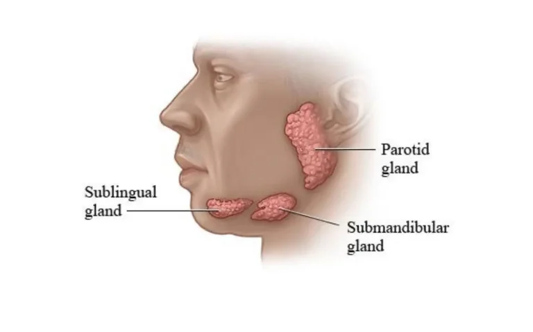
A salivary gland tumour is a lump that develops in the cells of the salivary glands. There are three pairs of major salivary glands: the parotid in front of the ear, the submandibular under the jaw and the sublingual under the tongue. In addition, there are hundreds of small (minor) salivary glands in the lining of the mouth, lips and throat.
Salivary gland tumours make up about 3–4 per cent of all head and neck tumours. About 80 per cent occur in the parotid gland, and about 80 per cent of parotid tumours are benign. In the submandibular gland under the jaw, the likelihood of a tumour being malignant is higher (about 25–50 per cent).
What are the symptoms?
The most common symptom is a painless swelling noticed in front of the ear, at the angle of the jaw or under the jaw. The swelling usually grows slowly; sometimes it is found by chance on an ultrasound or CT scan done for another reason.
- A swelling or lump in front of the ear, at the angle of the jaw or under the jaw
- A lump inside the mouth or on the palate
- Weakness on one side of the face, difficulty closing the eye or smiling
- Pain in the lump, rapid growth or attachment to the skin
- Numbness in the face or tongue, difficulty swallowing
Rapid growth, pain, facial weakness or numbness can suggest a malignant tumour; with these symptoms, assessment should not be delayed.
Risk factors
- Older age (it can occur at any age)
- Previous radiotherapy to the head and neck
- Smoking: closely linked in particular with Warthin’s tumour
- Certain occupational exposures, such as rubber manufacturing and nickel
How is it diagnosed?
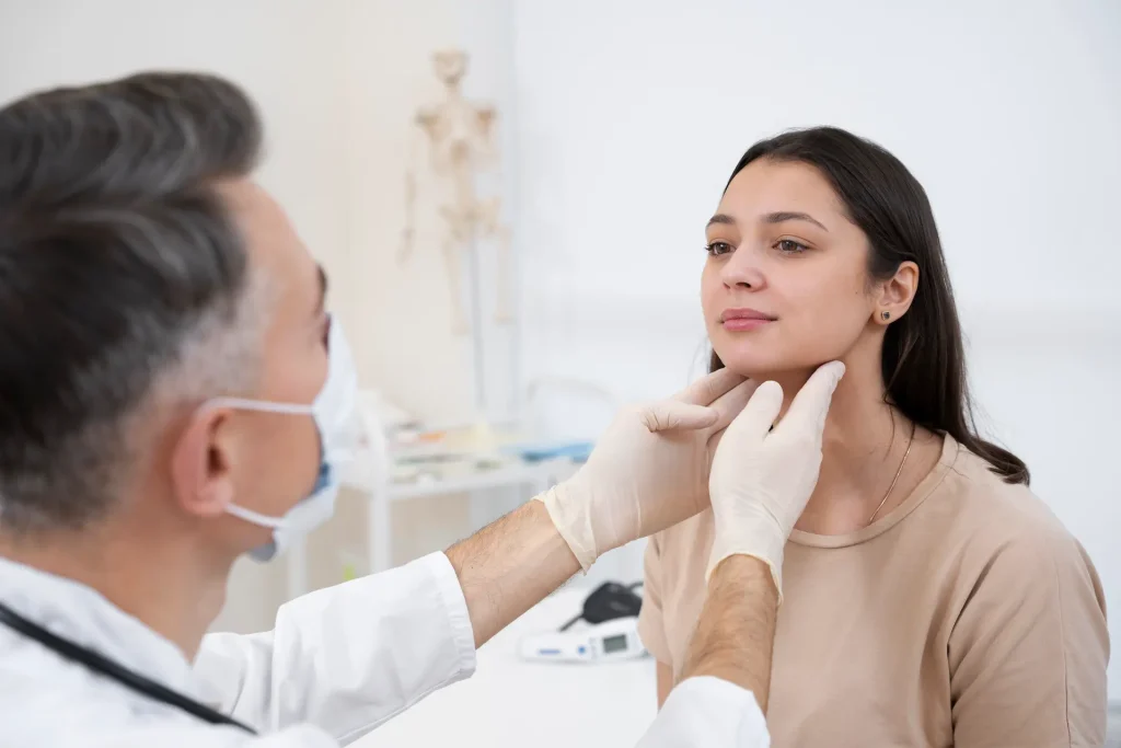
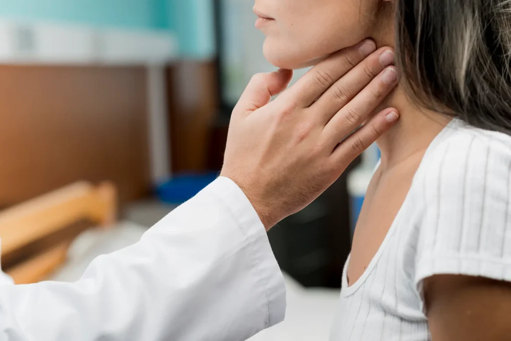
- Examination: the location, firmness and mobility of the lump and the function of the facial nerve are assessed; the inside of the mouth and the neck are examined.
- Imaging: ultrasound and, if needed, MRI or CT show the size of the tumour, which part of the gland it is in and whether it has spread.
- Fine-needle biopsy: a sample of cells is taken from the lump with a fine needle.
A needle biopsy does not always show the exact type of tumour. That is why, in some patients, the type is determined by a rapid pathology examination during surgery (frozen section) and by the final pathology after surgery; this information helps decide the extent of the operation.
Types of tumour
The World Health Organization classification describes more than 30 benign and malignant subtypes of salivary gland tumour. Knowing the type determines the extent of treatment and follow-up.
| Type | In brief |
|---|---|
| Pleomorphic adenoma | The most common salivary gland tumour; it is benign. Removal is recommended because, if left untreated for many years, it can rarely turn malignant. |
| Warthin’s tumour | Benign and found almost exclusively in the parotid; it is closely linked with smoking and occurs on both sides in about 10 per cent of cases. |
| Other adenomas | Slow-growing benign tumours such as basal cell adenoma and oncocytoma. |
| Mucoepidermoid carcinoma | The most common malignant salivary gland tumour; it has low-, intermediate- and high-grade types. |
| Adenoid cystic carcinoma | Seen more often in the minor salivary glands and the submandibular gland; it progresses slowly but tends to spread along nerves. |
| Acinic cell carcinoma | A low-grade cancer seen mostly in the parotid. |
How is it treated?
The first treatment for salivary gland tumours is surgical removal of the tumour together with the surrounding healthy tissue. For benign tumours, complete removal of the tumour is usually sufficient. For malignant tumours, depending on the type, grade and stage of the tumour, the lymph nodes in the neck may also be removed; radiotherapy may be added after surgery, and chemotherapy in advanced stages.
How is parotid surgery performed?
The facial nerve, which moves the muscles of the face, runs through the parotid gland and divides it into a superficial and a deep part. The most important aim of the operation is to remove the tumour completely while preserving the facial nerve and its branches.
- Superficial parotidectomy: if the tumour is in the superficial part above the nerve, this part of the gland is removed together with the tumour; the deep part continues to work.
- Total parotidectomy: for large, deep-lying or malignant tumours, the whole gland is removed.
- Extended surgery: if the cancer has spread to the surrounding tissues, these are also removed; a neck dissection is performed if needed.
The incision follows the natural skin creases in front of the ear and in the neck. During the operation, the facial nerve can be checked with electrical nerve monitoring (EMG). For tumours of the submandibular gland under the jaw, the gland is removed together with the tumour through an incision under the jaw.
After surgery: risk of facial palsy and recovery
After surgery, patients usually stay in hospital for two days and are discharged once the drain placed in the surgical area has been removed. In the first week the head is kept raised and the incision is kept dry; dressings and check-ups are done at the clinic.
Even when the facial nerve is preserved, temporary facial weakness can occur because the nerve is affected during the operation; in most patients this improves within weeks or months. Permanent facial palsy is rare; if the tumour has surrounded the nerve, or in malignant tumours, part of the nerve may need to be removed. This possibility is discussed openly before surgery.
Other risks
- Bleeding and a collection of blood under the skin (haematoma), infection
- Numbness of the earlobe and the surrounding area
- Frey’s syndrome: sweating or redness of the skin over the surgical area when eating
- A hollow or scar in the surgical area
- In submandibular gland surgery, effects on the nerves that move the tongue or the corner of the mouth
Outcomes and follow-up
Benign tumours are mostly treated successfully with surgery, and the recurrence rate is low. In malignant tumours, the outcome depends on the type, grade and stage of the tumour; long-term survival rates are high for tumours caught at an early stage and for low-grade tumours.
After treatment, follow-up is carried out with regular examinations and, where needed, imaging.
How does the process work with Prof. Dr. Öner Çelik?
- Consultation: at the clinic in Suadiye, the lump and the facial nerve are assessed; any previous ultrasound, MRI and biopsy results are reviewed.
- Tests and plan: any missing tests are completed; the extent of the operation, the risks related to the facial nerve and the recovery process are discussed.
- Surgery: performed at Maltepe University Hospital.
- Follow-up: dressings, check-ups and any further treatment plan based on the pathology result are handled at the clinic; where needed, decisions are made together with radiation oncology.
Details are on the your treatment in Istanbul page.
Frequently asked
Could a swelling in front of the ear be a salivary gland tumour?
It could. Salivary gland tumours are most often found in the parotid gland in front of the ear, and most are benign. The diagnosis is made by examination, ultrasound or MRI and fine-needle biopsy; the main treatment is surgical removal of the tumour while preserving the facial nerve.
More detail: Salivary gland tumours.
Are salivary gland tumours painful?
Salivary gland tumours are usually noticed as a painless swelling. Pain in the lump, rapid growth or weakness in the face can suggest a malignant tumour and should be assessed without delay.
Can parotid surgery cause facial paralysis?
Temporary facial weakness can occur after the operation and improves within weeks or months in most patients. Permanent facial paralysis is rare; the risk is higher when the tumour surrounds the nerve and is discussed openly before the operation.
Is every salivary gland tumour cancer?
No. Most salivary gland tumours, especially those in the parotid gland, are benign. Ultrasound or MRI and fine-needle biopsy are used to identify the type; the definitive diagnosis is made by pathology.
Articles on this topic
- Salivary Gland Tumours and Surgery: Is There a Risk of Facial Paralysis? After salivary gland surgery, particularly parotid surgery, temporary weakness of the facial nerve can occur; it usually recovers…
References
- WHO Classification of Tumours Editorial Board. Head and Neck Tumours. 5th ed. Lyon: IARC; 2022.
- NCCN Clinical Practice Guidelines in Oncology: Head and Neck Cancers (Salivary Gland Tumors).
This content is for information only and does not replace medical advice. Every medical procedure requires an individual assessment and may carry risks. Results vary from person to person.