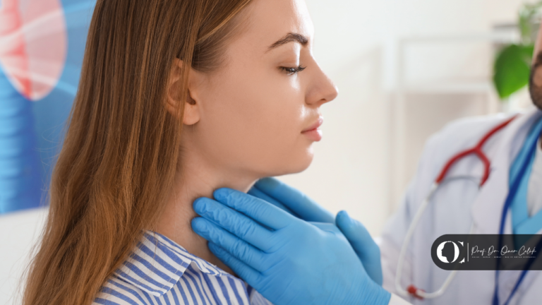


You have noticed a firm lump just in front of your earlobe, at the angle of your jaw or under your tongue. Perhaps it has been there for weeks, perhaps you have only just noticed it. When you search for “swelling in front of the ear”, one of the first results is salivary gland tumour. And right after it comes the big fear: “If I have surgery, will my face be paralysed?”
I am Prof. Dr. Öner Çelik. As an ear, nose and throat and head and neck surgeon, I have seen the deep worry this question creates in the eyes of many patients throughout my career. Our face is our identity: our smile, our gaze, our expression. So it is entirely human to hesitate when surgery is mentioned in an area so close to the facial nerve.
Today we draw back that curtain of fear. What are salivary gland tumours, how is parotid surgery performed, and with today’s surgical technology, how manageable is the much-feared risk of “facial paralysis”? Let us look at the subject carefully and in detail.

Where Are Our Salivary Glands?
We have three pairs of major salivary glands:
-
Parotid gland: the largest salivary gland, located in front of and below the earlobe. 80% of tumours arise here.
-
Submandibular gland: located just below the jawbone.
-
Sublingual gland: located under the tongue.
There are also hundreds of small (minor) salivary glands inside the mouth, on the palate and in the cheeks. When it comes to salivary gland tumours, the focus of surgeons and patients is usually the parotid gland. Why? Because the vital line that controls all the movements of the face runs through it: the facial nerve.
The Facial Nerve: A Hidden Path Inside the Gland
You can think of the parotid gland as a sandwich. The facial nerve runs right through the middle of it and divides the gland tissue into a “superficial” and a “deep” part. This nerve is the main electrical cable that lets us close our eyes, smile, raise our eyebrows and move our lips.
The real skill in salivary gland tumour surgery (parotidectomy) is not simply removing the tumour, but separating it from the tissue while preserving, one by one, the hair-thin nerve branches that run beside, beneath or above it. That is why this operation is an art of microsurgery.
What Are the Symptoms of a Salivary Gland Tumour?
Salivary gland tumours are usually “silent” guests. They do not cause pain and grow insidiously. Patients usually come with the following complaints:
-
A slowly growing, painless firm lump in front of the ear or under the jaw.
-
An increase in swelling while eating (in some cases).
-
If the tumour has grown very large and is malignant (cancer): reduced movement or numbness on that side of the face.
So why do so many people ignore these swellings? Because when there is no pain, we tend to assume the problem is not serious. Yet with salivary gland masses, early diagnosis is everything when it comes to preserving the nerve.
Benign or Malignant?
About 80% of salivary gland tumours are benign. The type we see most often is pleomorphic adenoma. Although this tumour is benign, if left alone it can grow very large over the years and, rarely, turn into cancer (carcinoma ex pleomorphic adenoma). In addition, as the mass grows, the pressure it puts on the facial nerve increases and makes the operation more difficult.
The other common benign type is Warthin’s tumour, which we see particularly in men who smoke.
Malignant tumours grow faster, are sometimes painful and tend to involve the facial nerve. Mucoepidermoid carcinoma and adenoid cystic carcinoma are the cancer types we encounter most often.
Diagnosis: The Right Path, Step by Step
How does the process work once a mass is noticed?
-
Physical examination: the firmness and mobility of the mass are checked.
-
Ultrasound and MRI: the gold standard for seeing the borders of the mass and its relationship to the facial nerve.
-
Fine-needle aspiration biopsy (FNAB): a fine needle is inserted into the mass to take a cell sample. This gives us vital information for planning the operation (benign or malignant?). Don’t worry: needle biopsy does not cause the tumour to spread; on the contrary, it lights the surgeon’s way.
How Is Parotid Surgery (Parotidectomy) Performed?
Once surgery has been decided, what patients most want to know are the technical details. The operation is performed under general anaesthesia, usually through a cosmetically planned incision that starts in front of the ear and extends below the jaw, designed to sit within the natural neck creases as it heals.
Surgical techniques:
-
Superficial parotidectomy: if the tumour is in the tissue above the nerve, only this part is removed.
-
Total parotidectomy: if the tumour is in the deep tissue below the nerve or is malignant, the whole gland is removed.
The most critical moment of the operation is finding the main trunk of the facial nerve. Once the nerve has been found, the tumour is “peeled” away from it millimetre by millimetre.
The Big Question: What Is the Real Risk of Facial Paralysis?
Now to the much-feared topic. In modern surgery we have two main tools to protect the facial nerve: careful surgical technique and technology.
1. Nerve Monitoring (NIM Device)
Today we use devices called “nerve monitors” during parotid surgery. Thanks to electrodes placed on your facial muscles during the operation, the device gives an audible warning when we get close to the nerve. It is like a “radar” in the surgeon’s hand on a dark road. It helps us confirm the position of the nerve and protect even the smallest branches.
2. Temporary vs. Permanent Paralysis
-
Temporary weakness: when the tumour is being separated, touching or slightly stretching the nerve can cause it to be “stunned” (neurapraxia). In that case there may be temporary weakness at the corner of the mouth or in the eyelid after surgery. This usually recovers completely within 2–6 weeks (sometimes a few months).
-
Permanent paralysis: if the tumour is not very large and is not an aggressive cancer directly invading the nerve, the risk of permanent facial paralysis is below 1–2%.
“Our task is not only to remove the mass, but to restore the patient’s health while doing everything we can to preserve their smile and expression.”
After Surgery and Frey Syndrome
After the operation, patients usually stay in hospital for one night. A small “drain” (tube) is placed to remove any fluid that may collect in the area and is taken out the next day.
What is Frey syndrome? In some patients, months after surgery, the skin over the operated area may sweat or flush while eating. This is called Frey syndrome. It happens because nerve fibres that used to go to the salivary gland mistakenly connect to the sweat glands as they heal. It is usually not a serious problem, and treatment is available if needed.
Frequently Asked Questions
1. Can a salivary gland tumour be cured with medication? Unfortunately not. Salivary gland tumours are anatomical growths, and the treatment is surgical removal.
2. Will the scar be very visible? The incision is placed in the natural folds of the ear and the lines of the neck, so once healing is complete it is usually difficult to notice from the outside.
3. What happens if I don’t have surgery? Even if it is benign, the tumour keeps growing. As it grows, it presses more on the facial nerve and pushes it aside, which increases the risk considerably if you have surgery later. There is also a small risk of it turning into cancer.
4. Will I have less saliva after surgery? Usually not. Because the body has many other salivary glands, removing part or all of one gland does not usually cause a dry mouth.
5. What should I pay attention to after parotid surgery? For the first few weeks you should avoid strenuous exercise, protect the wound from the sun and carry out massage and dressing care as your doctor recommends.
Conclusion: Overcoming Fear with Knowledge
Being diagnosed with a salivary gland tumour can be frightening, but you should know that head and neck surgery has advanced considerably in this field. Thanks to technology (nerve monitors), microscopic magnification techniques and cosmetic approaches, these operations can now be performed safely.
The smile on your face matters greatly to us. If you have noticed a firm lump in front of your ear, do not neglect yourself out of fear of paralysis. Remember that treating a small mass at an early stage usually makes it much easier to preserve the nerve.
I wish you healthy, peaceful days full of smiles.
References
- Sood AJ et al. Facial nerve monitoring during parotidectomy: a systematic review and meta-analysis. Otolaryngol Head Neck Surg, 2015. pubmed.ncbi.nlm.nih.gov
- PDQ Adult Treatment Editorial Board. Salivary Gland Cancer Treatment (PDQ): Patient Version. National Cancer Institute. pubmed.ncbi.nlm.nih.gov
This content is for information only and does not replace medical advice. Every medical procedure requires an individual assessment and may carry risks. Results vary from person to person.